
Anatomical Landmarks of the Mouth AmberaresKing
A thorough knowledge of oral anatomy helps the clinician in identifying enough landmarks that in turn act as positive guides in treatment planning. In the present article, a review of all the intraoral anatomical landmarks is been presented and analyzed Keywords: Maxillary ridge, Mandibular ridge, Edentulism, Anatomical landmarks
landmarks of face and oral cavity
The stomatognathic system includes various anatomical structures, which allow the mouth to open, swallow, breathe, phonate, suck and perform different facial expressions. These structures are the temporomandibular joint (TMJ), jaw and mandible, muscle tissues and tendons, dental arches, salivary glands, as well as the hyoid bone and the muscles that connect the latter to the scapula and the.

Surrounding Muscles of Upper Complete Denture. Dentistry, Dental hygiene student, Dental
Revisions: 15 format_list_bulleted Contents add The oral cavity, better known as the mouth, is the start of the alimentary canal. It has three major functions: Digestion - receives food, preparing it for digestion in the stomach and small intestine. Communication - modifies the sound produced in the larynx to create a range of sounds.

Oral cavity anatomy with educational labeled structure vector illustration
We have created 110 medical original illustrations of the mouth, the buccal cavity, the bones of the palate, the tongue, the salivary glands and the oral part of the pharynx with vessels and nerves.

mandibular structures Radiographic Anatomy Pinterest Anatomy, Dental and Dental anatomy
Hand: Anatomy, is a complex. Diagram of the mouth and lips showing their different components and landmarks. Image: "The mouth includes the lips, tongue, palate, gums, and teeth" by OpenStax College. License: CC BY 4.0 Movement. Numerous muscles are responsible for movement of the lips.
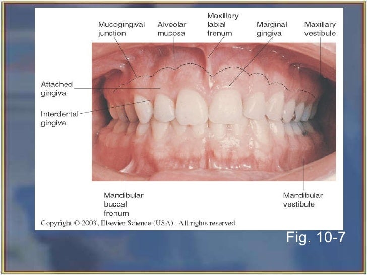
landmarks found in the oral cavity
The anatomical landmarks of the oral fissure and the lips in a 20-year-old female. Full size image. The lips (labialis, superioris,. It contributes to the modiolus of the mouth in addition to the other facial muscles contributing to the tendinous chiasm lying slightly superior to the lateral commissures of the oral fissure (Hur et al. 2010a, b).

Anatomical Landmarks of Edentulous Jaw. Dental hygiene student, Dental assistant study, Dental
The cavity is separated into anterior and posterior parts by the dental arches (or teeth): the anterior oral vestibule sits anteriorly to the teeth and behind the lips, whilst the oral cavity proper describes the area behind the teeth.

Mouth Teeth Diagram with Label coordstudenti
The easiest anatomical landmark in the floor of the mouth examination, is the curvature of the tongue's surface. Also important is the determination of the air-tissue interface [3, 6]. Intraoral probes are more useful when we want to examine the tongues surface since they are easier to handle in such a small region.

Maxillary Landmarks Labial frenum, Incisive papilla, Buccal frenum, Maxillary alveolar ridge
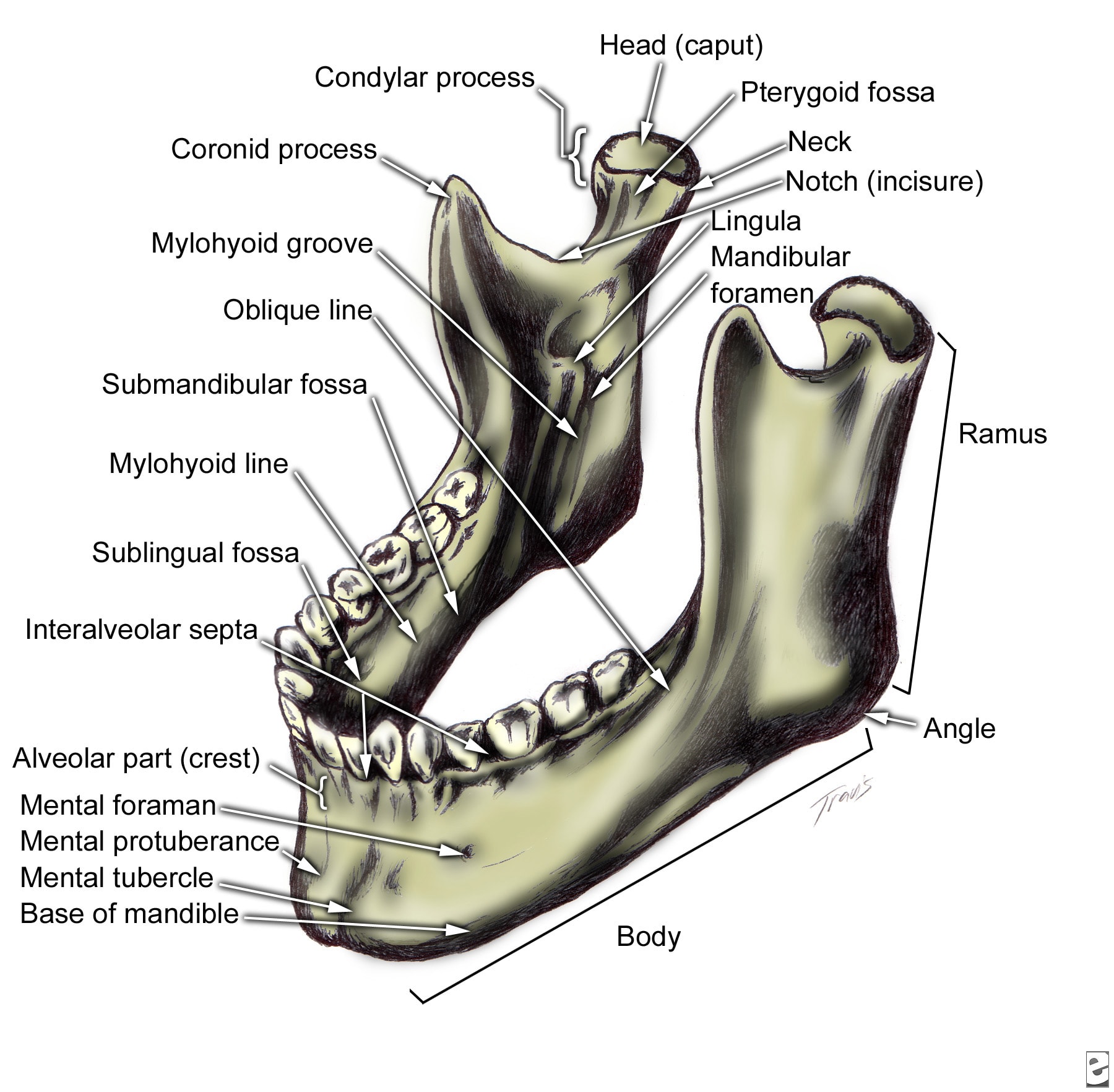
The base is the inferior part of the body that features several anatomical landmarks. On its external surface, we can identify: . The mandibular symphysis: Fibrous tissue in the midline of the mandibular body, which ossifies by the first year of life.It unites the left and right halves of the mandible in order to form a single, symmetrical bone. The mental protuberance: A bony prominence at.

Facial landmarks divided into anatomical and pseudoanatomical... Download Scientific Diagram
Figure 2 below, includes many of the normal anatomical landmarks that will be visible on a diagnostic panoramic image. The maxillary sinuses are radiolucent and can be found bilaterally on either side of the nasal septum. The zygomatic process is a vertical, radiopaque line that forms the anterior portion of the zygomatic arch (cheekbone)..

Anatomical Landmarks of the Mouth EennHorton
Normal Anatomic Landmarks of the Head Neck and Oral Cavity Bone Structure of the Face Facial Landmarks Landmarks in the Oral Cavity Teeth in the Oral Cavity Types of Teeth, Structures, Location and Functions Divisions and Components of the Teeth Types of Teeth and their Functions Surfaces of the Teeth Dentitions Primary Dentition
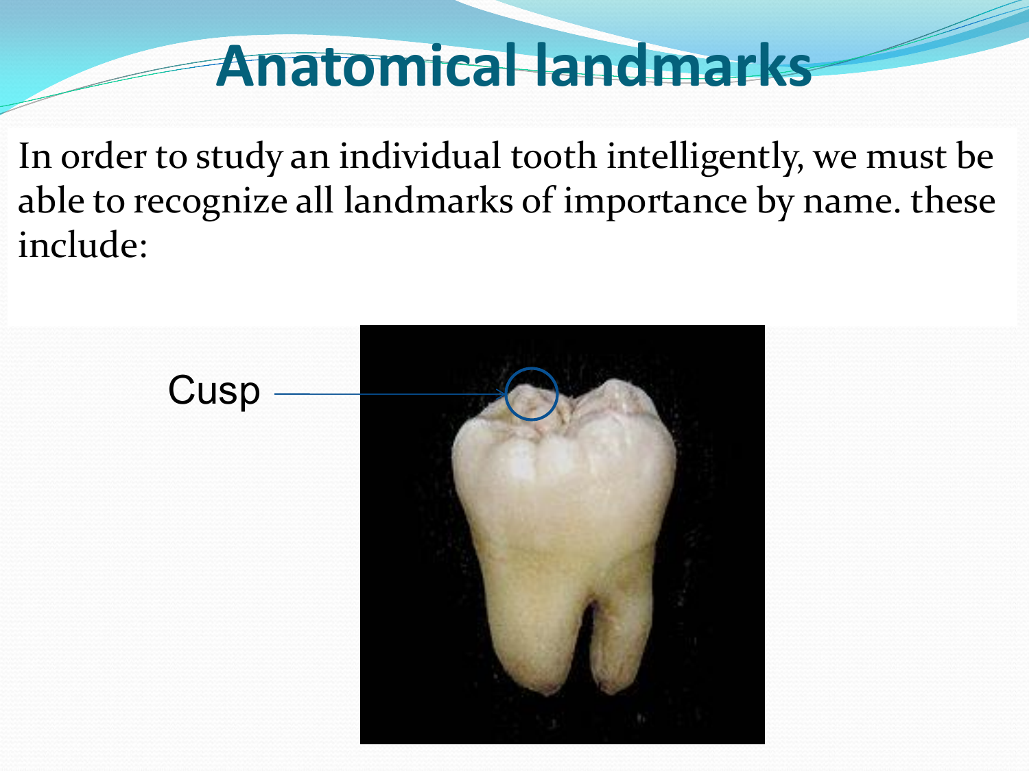
Anatomical landmarks
1. Describe the basic anatomy of the ear, nose, mouth, and throat. 2. Perform a basic examination of the ear, nose, mouth, and throat, identifying normal and pathological conditions. 3. Properly use an otoscope to examine the ear and the nose. 4.

Anatomical landmarks in mandibular edentulous arch YouTube
These landmarks also form a benchmark for determining normal facial anatomy when performing an extraoral examination on a patient. 1 Figure 2. Facial Landmarks. Ala - Wing of the nose. Inner canthus of the eye - The inner corner of the eye. Labial commissures - Corners of the mouth.
vestibule anatomy mouth
Landmarks of the oral tissues include the palate, tongue, cheeks and floor of the mouth. It is significant to recognize the normal appearance of these structures during an intraoral examination of the patient. Fauces - Passageway from oral cavity to pharynx.

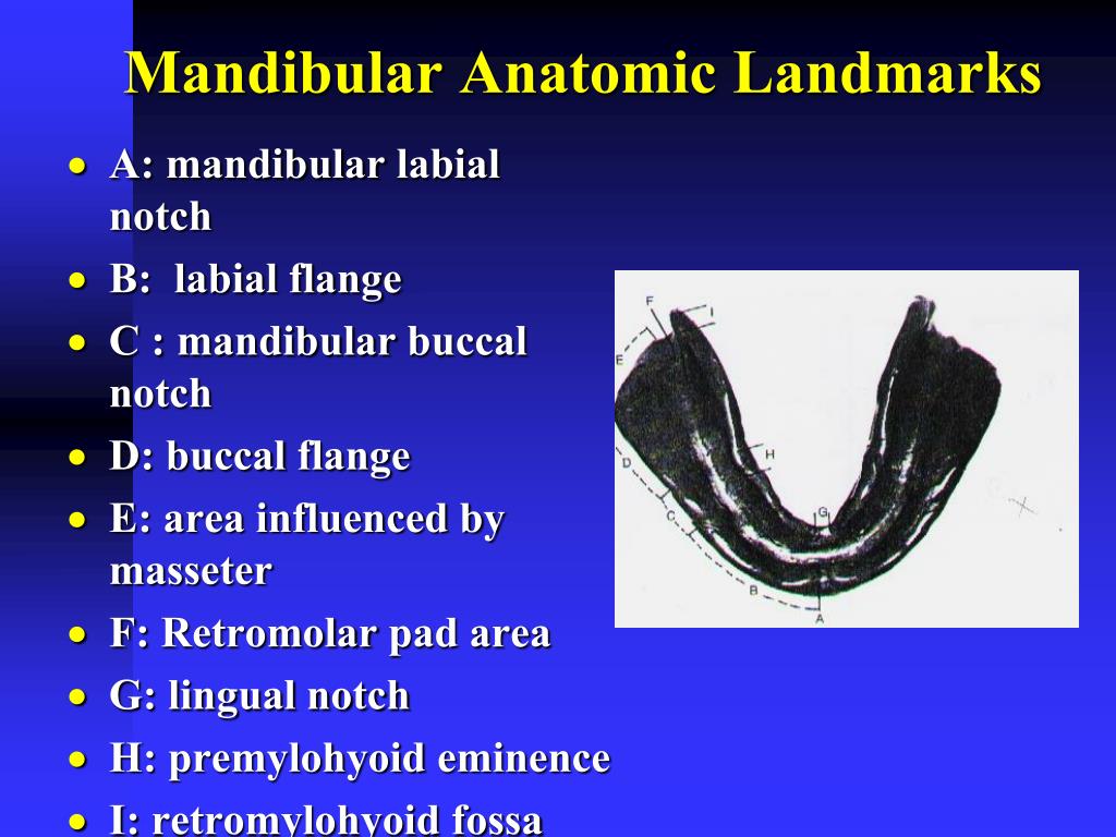
Anatomic Landmarks Dentures Mouth
The tooth is one of the most individual and complex anatomical as well as histological structures in the body. The tissue composition of a tooth is only found within the oral cavity and is limited to the dental structures. Each tooth is paired within the same jaw, while the opposing jaw has teeth that are classified within the same category. However they are not grouped according to structure.
ORAL & MAXILLOFACIAL SURGERY Facial Bone Anatomy
The modiolus is the anatomical point at the corner of the mouth or angle of the mouth where the orbicularis oris, buccinator, caninus, triangularis, and zygomaticus muscles intersect and are located near the rim of the mouth (Fig. 6.44). It is also called as the fibromuscular condensation where the extrinsic and intrinsic muscles meet together.